The Method
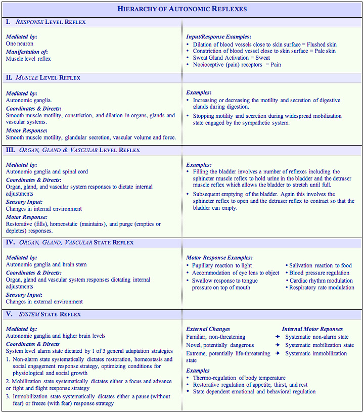
Hierarchy of Autonomic Reflexes
Autonomic reflexes are moderated and coordinated in a hierarchical, integrated fashion by three subsystems of the autonomic nervous system:
- The parasympathetic system,
- The sympathetic system, and
- The system managed by the non-myelinated vagus nerve
The autonomic system can engage simple responses at the local level, involving only part of one neuron and at the regional muscle level, coordinated and directed (mediated) only by the autonomic ganglia; or engage progressively more complex autonomic reflexes as the autonomic ganglia work in conjunction with the central nervous system at the spinal cord level, brain stem level, and higher brain levels to coordinate and direct more complex reflex responses. In general, the higher the level of complexity, the more likely the reflex will require coordination of not only the sympathetic and parasympathetic responses, but with somatic responses as well. (R. Rhoades, D. Bell, Medical Physiology, Lippincott, Williams & Wilkins, 2009, Third Edition, p. 118). The following summary characterizes in more detail the hierarchical levels of autonomic reflexes.

Parasympathetic & Sympathetic Tone & the Symbiotic Nature of Control
Under normal conditions, the sympathetic and parasympathetic systems both exert continuous influence on the local organs, glands and vascular systems they manage. This continuous level of control is referred to as tone. The tone of each of these systems is based on the relative level of firing each is directing at the target area. Tone can range anywhere from high to low. High indicates dominant control while low indicates subordinate control. For example, when entering a dark room, the sympathetic neurons begin firing at a high rate toward the iris of the eye, causing it to dilate the pupil so that more light can enter while the firing of the parasympathetic nerve drops to very low levels. In this case sympathetic tone would be high, parasympathetic tone would be low and the sympathetic system would be considered dominant at the local level of the iris and pupil. When returning to brightly lit area parasympathetic neurons begin firing at a high rate causing the iris to constrict the pupil so that less light enters. Again, at the same time firing increases for the parasympathetic system, firing decreases for the sympathetic system. In this case, parasympathetic tone would be high, sympathetic tone low and the parasympathetic system would be in control at the local iris level.
While both systems work continuously at the localized level, only the sympathetic system has the capacity to produce widespread responses in the body. The body when faced with novel, potentially dangerous changes in the environment triggers widespread control. When this occurs, the body adapts what is called a mobilization strategy, readying the body to respond quickly and efficiently to deal with the pending challenge. This state can be engaged when faced with challenging tasks mobilizing the body to focus and advance to complete the task or when face with real danger mobilizing the body for fight or flight. Regardless of reason, for mobilization, the sympathetic system takes over on a widespread, global basis slowing or shutting down unnecessary processes to recruit and allocate the body’s resources to mobilize. The following chart summarizes how the parasympathetic and sympathetic systems influence a cross section of organs, glands and vascular systems when each is in dominate control. Click on the body part impacted (internal effector) if you would like to know more about its function.
|
Autonomic System |
Parasympathetic subsystemNon-Alarm State Response |
Sympathetic subsystem |
|---|---|---|
| Eyes | ||
| Pupil & Iris |
Constricts (narrows) pupil as the inner muscles of the iris contract to let in less light and to make it easier to view nearby objects. |
Dilates (widens)pupil as the outer muscles of the iris contract to let in more light and heightens the body’s visual awareness. |
| Ciliary muscle |
Contracts ciliary muscle to allow the eye’s lens to focus on nearby objects. |
Relaxes ciliary muscle to allow the eye’s lens to focus on distant objects. |
| Lacrimal gland | Releases tears | None |
| Nasal glands | Produces mucus | Inhibits mucus |
| Salivary glands |
Secretes larger volume of thin, watery saliva rich in enzymes and low in protein. |
Secretessmaller amounts of saliva rich in proteins. |
| Repiration | ||
| Trachea | Constricts trachea tube | Keeps trachea open |
| Bronchial Tubes | Contractssmooth muscle, causing bronchial tubes and branches to constrict, allowing less air in lungs. | Relaxessmooth muscle, causing bronchial tubes and branches to dilate, allowing more air in lungs, and making more oxygen available for muscle contraction that may be needed for mobilization. |
| Bronchial Glands | Normalizes Secretion | Increases or decreases secretion based on pharmacological agent present in system. |
| Blood vessels | ||
| In Lungs | None | Dilates blood vessels |
| In Skin | None | Constricts to send more blood to major muscle groups for mobilization |
| In remaining trunk & limbs | None | Dilates blood vessels |
| Heart | Decreases heart rate and force of contraction. | Increases heart rate and force of contraction. |
| Adrenal Glands | None |
Releases stress hormones |
| Liver | Stores glucose to be used by body when needed. | Releases glucose |
| Pancreas | Secretes insulin and enzymes |
Inhibits insulin secretion |
| Kidneys | Normalizes urine output |
Decreases urine output |
| Digestion | ||
| Stomach | Increases production and secretion of digestive enzymes. |
Decreases production and secretion of digestive enzymes. |
| Intestines | Normalizes based on need |
Slows peristalsis, muscular action of the intestines that keeps food moving through the intestines. |
| Intestinal glands | Normalizessecretion |
Inhibits secretions |
| Skin | ||
| Sweat glands | None |
Increases sweat production to cool the body during mobilization |
| Piliorector muscles | None |
Contracts when cold or scared. For human ancestors this provided puffed out hair/fur trapped body heat and created an additional level of insulation and made them appear bigger and more threatening when mobilizing to face danger. |
| Facial & Neck Muscles | Dilates |
Constricts |
| Skeletal Muscles | None |
Dilates or constricts depending on pharmacological agents in the body |
| Urinary System | ||
| Ureter | Relaxes |
Contracts |
| Detruser | Contracts |
Relaxes |
| Sphincter | Relaxes |
Contracts |


